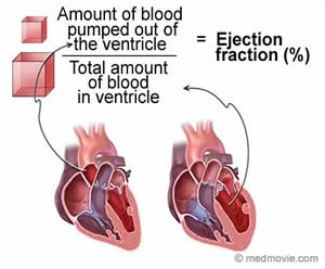
The ejection fraction is the amount that doctors use to calculate the percentage of blood that comes out of your heart each time you inhale.
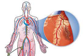
As your heart beats, it pumps (pumps) blood out of your body from the two lower extremities, known as the left and right ventricles. During the beating, when your heart is relaxed, two ventricles fill with blood.
However, it takes more than one suction to pump all the blood into the ventricle. Part of the ejection test your doctor can use to determine the percentage of blood that comes out of the left ventricle each time your heart beats, as well as to understand how well your heart is working.
The ejection fraction can help diagnose heart failure. Cardiac design, with left and right ventricles in the lower part of the heart.
How is the ejection fraction measured?
Generally, your left ventricle is the measured portion of the ejection. It carries heavily to your body, pumping blood to almost every major organ in your body. However, current research suggests that the right ventricle should not be overlooked when determining the right portion of the right heart ejection. The accuracy of left ventricle ejection fraction (LVEF) can be measured using a variety of imaging techniques. The most common steps to check the partial ejection include:
Echocardiogram– An echocardiogram uses sound waves to take pictures of your heart. A 2012 research article suggests that 3-D images provide excellent reading and accuracy.

Cardiac MRI (C-MRI)-C-MRI scans are based on imaging using a magnetic field, radio waves, and a computer to create detailed images of your heart.

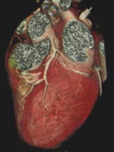
Cardiac catheterization– In this procedure, your doctor inserts an empty tube into a large artery to monitor your heart rate. During catheterization, coronary angiography was performed. The dye is injected into a catheter. Then, an X-ray examines the blood flow throughout your heart.
Cardiovascular scanner for heart-Tracking the amount of radioactive material injected into your bloodstream. They are then detected by cameras that produce images of your heart and its ways.
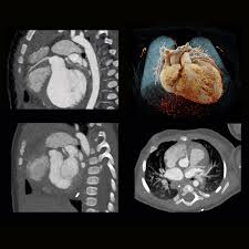
Cardiac CT scan-This X-ray procedure can provide a representation of the size of the heart and, with images attached to the gate, the function of the heart.
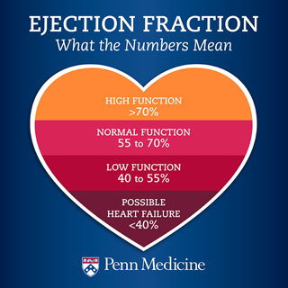
What do ejaculation results mean?
According to a reliable heart specialist, the results of LVEF are usually classified as follows for people 18 years and older:
LVEF lists Men as Women
Standard range 52–72% 54–74%
Minimum width 41–51% 41–53%
Unusual range 30–40% 30–40%
The extraordinary range of less than 30% less than 30%
The a very high number of LVEFs. An LVEF level of more than 72% of men and more than 74 percent of women may be indicative of a condition such as hypertrophic cardiomyopathy. This is when the heart muscle becomes larger than normal and disrupts cardiac function. It is a common cause of sudden cardiac arrest.
Normal. Even if you have a normal LVEF dose, it does not necessarily mean that your heart is “normal.” Some people may have heart failure with a condition known as heart failure with proven ejection fraction (HFpEF).
Slightly lowered. The ejection rate between 41 and 51 percent in males and between 41 and 53 percent in females is classified as slightly reduced. It could be a sign of a heart attack, perhaps due to a heart condition or a previous heart disease.
A sign of heart failure. LVEF is less than 40 percent heart failure with reduced ejection fraction (HFrEF). It may also be due to cardiomyopathy, in which your heart muscle weakens, making your heart less efficient in pumping blood throughout your body.
What are the types of heart failure?
Heart failure with decreased left ventricular function (HFrEF)
An ejection fraction of less than 40 percent is classified as a cardiac failure with reduced ejection fraction (HFrEF). It happens when one of the chambers of your heart cannot enter properly. Medications can cure it.
Symptoms may include:
- Shortness of breath
- Fatigue
- Heartbeat
- Dizziness, confusion, lightheadedness
- Swelling in the ankles, legs, or abdomen
- Show intolerance
You may also be at greater risk for an abnormal heartbeat which can be dangerous to health.
If your ejaculation rate is below 35 percent, your doctor will likely recommend other treatments, such as an implanted cardioverter defibrillator or pacemaker, to help control your heart rate.
Cardiac failure by preserved left ventricular function (HFpEF)
With this type of heart failure, you have a stored, or normal, part of the discharge. Occurs when your left ventricle is not properly rested.
This may be due to the hardening of the heart muscle or the stiffness of the heart muscle. It can cause small amounts of blood to be pumped from the heart to the rest of your body.
Symptoms may include fatigue and shortness of breath during exercise. HFpEF can be the result of aging, diabetes, or high blood pressure.
What can cause a reduced ejection fraction?
As we grow older, our hearts also grow. The walls of the heart thicken and lose their ability to wrap and relax properly.
But studies of the lower part of the ejection may indicate other types of heart damage, including:
- Cardiomyopathy– Cardiomyopathy is a breakdown of the heart muscle caused by tightening or opening of the heart muscle. This makes it difficult for your heart to pump properly.
- Heart disease and coronary artery disease-A heart attack occurs when one or more of your arteries close, causing heart muscle damage. Coronary artery disease can narrow or block your left and right arteries, making it more difficult for blood to flow to your heart.